A validated ladder that simplifies agarose gel electrophoresis size estimation with consistent, reproducible results.
| Number | Specifications | Catalog Price | Add To Cart |
|---|---|---|---|
| YD19161 | 2×500µl | Online Consult | + |
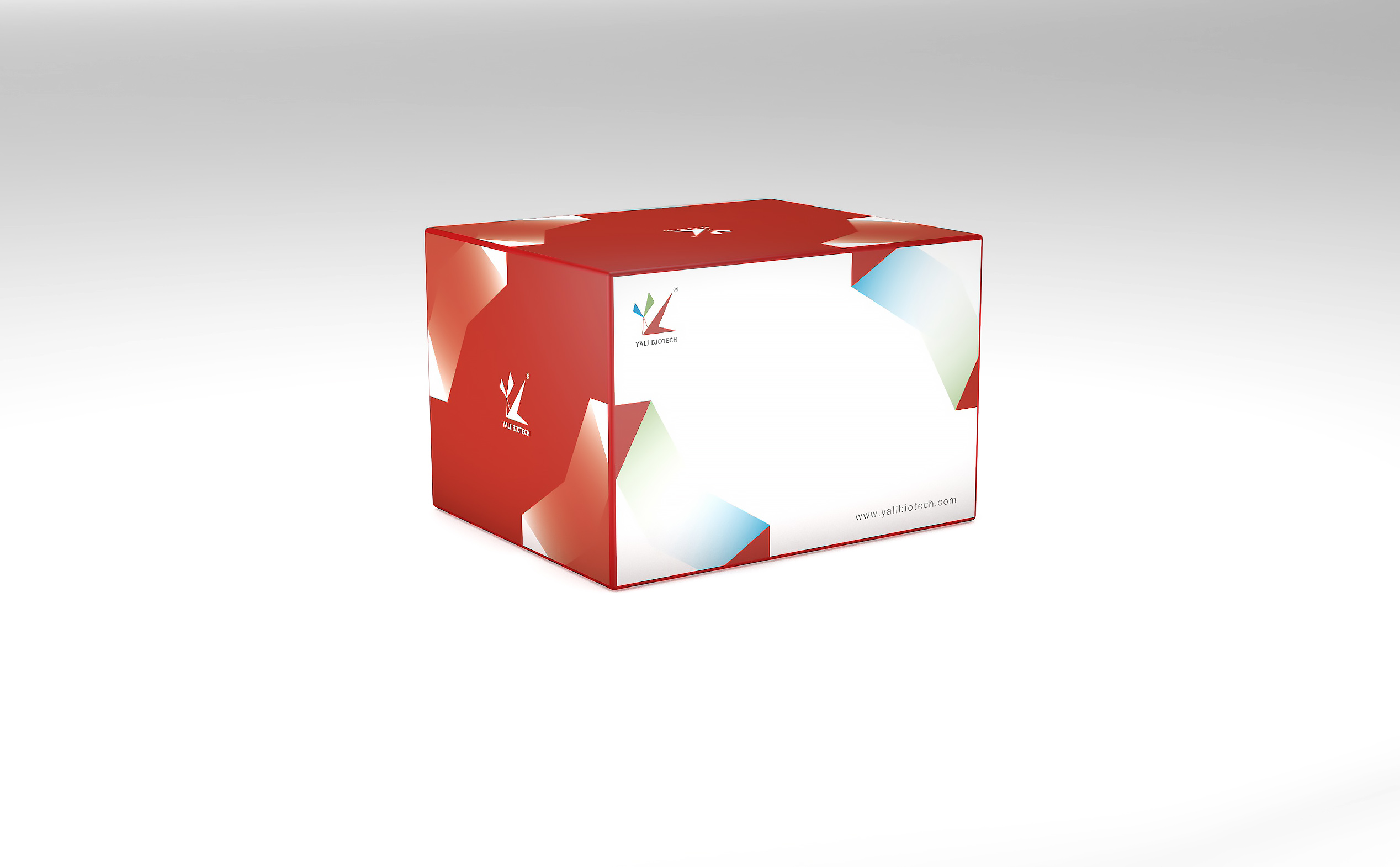
YALEPIC® 15K DNA Marker consists of 11 double-stranded DNA fragments of specific molecular weight and is mixed with 1×loading buffer containing a blue dye, which can be used as standard reference in agarose gel electrophoresis. All the fragments in the DNA Marker/Ladder are obtained by enzymatic digestion and purification. The bands are more clear during electrophoresis and the mass ratio between the bands is more accurate. The 500 bp fragment at concentration of 100 ng/5 μl, which are bright bands; 1500bp fragment at concentration of 75 ng/5 μl;all the remaining fragments are at a concentration of 40~50 ng/5 μl.
Bands:100bp,250bp,500bp,750bp,1000bp,1500bp,2000bp,3000bp,5000bp,8000bp,15000bp.
Store at -30 ~ -15°C for 3 years,at 2~8 ℃ for one year; transport at -20 ~ 30℃.
Not recommended for RNA molecular weight determination. Double-stranded DNA in the Marker maintains its double-stranded structure in standard agarose gel electrophoresis, while RNA migrates differently than DNA of the same size, leading to inaccurate size estimation. For rough observation of RNA band distribution, it can be used temporarily, but not for precise quantification. For denaturing gel electrophoresis, DNA strands will denature and the Marker absolutely should not be used for RNA size indication.
Common causes: (1) Stale electrophoresis buffer – after multiple uses, ion strength decreases and pH rises, reducing buffering capacity; replace with fresh buffer each time. (2) Nuclease contamination – use sterilized tips, tighten caps after use. (3) Gel quality issues – use high-quality agarose and ensure even solidification. (4) Excessive voltage – recommend 4-10 V/cm; higher voltage generates excess heat causing band diffusion.
This is a ready-to-use product containing 1x Loading Buffer – no additional addition or heating needed. Standard conditions: for well widths <6 mm, load 5 microL directly; for wider wells, increase to 8-10 microL. Recommended gel: 1%-2% agarose, voltage 4-10 V/cm, electrophoresis 25-60 minutes.
(1) More bands: 11 bands covering 100bp-15kb, vs 7-8 bands in most competitors, for higher resolution. (2) Dual indicator design: 500bp bright band + 1500bp secondary bright band for quick positioning and concentration estimation. (3) Ready-to-use: pre-mixed Loading Buffer, no preparation needed. (4) Concentration gradient design: differentiated band concentrations for semi-quantitative analysis.
2-8 degrees C for 2 years, -20 degrees C for 3 years. For frequent use, store at 2-8 degrees C; for long-term storage, use -20 degrees C. To avoid band degradation from repeated freeze-thaw, aliquot into small single-use portions after first use.
The 15K Marker covers 100bp-15kb, suitable for medium-to-large DNA fragment detection (plasmid digest analysis, large PCR product identification). For fragments 100bp-2kb, choose the 2K Marker for higher resolution. For fragments above 15kb, choose 10K or higher specification products. Select the product that best matches your target DNA fragment size for optimal resolution.
The product design has 500bp at 100 ng/5 microL (bright), 1500bp at 75 ng/5 microL (secondary bright), others at 40-50 ng/5 microL. This differentiated design aids quick positioning and semi-quantification. If unexpected unevenness occurs, check: accurate loading volume (5 microL recommended), appropriate gel concentration (1% agarose recommended), and fresh buffer.
Compatible. The YALEPIC 15K DNA Marker works with GelRed, SYBR Green, Goldview, and other common nucleic acid fluorescent dyes. Note: different dyes have varying staining efficiencies that may affect band brightness. When using a new dye type, optimize exposure parameters with a pilot experiment. For EB staining, use 0.5 microgram/mL EB solution for 15-20 minutes.
Normal product should be a clear, transparent solution. If precipitate is found, possible causes: (1) SDS precipitation from temperature fluctuations during transport; (2) Trace precipitate after long-term storage. Handling: place at room temperature or warm gently at 37 degrees C for 5-10 minutes, mix by gentle inversion. Do not vortex or boil, as this may denature DNA.
Typical applications: (1) PCR product fragment size verification; (2) Plasmid DNA restriction digest identification – confirming recombinant plasmid construction through digest fragment sizes; (3) Genomic DNA restriction fingerprinting – Southern blot pre-hybridization fragment size screening; (4) DNA fragment quality assessment before gel recovery – determining target band position for excision; (5) Ligation and digestion product fragment identification and semi-quantitative concentration analysis.




Production center: 4th floor, Building 9, Yangshan Science and Technology
Industrial Park,No. 8 Jinyan Road, Suzhou High-tech Zone
Marketing center: Pujiang High-tech Park, Minhang District, Shanghai
Copyright: 2026 YALI Biotech Co., Ltd.
 Scan to follow us